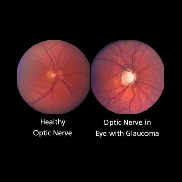
The optic nerve carries impulses for sight from the retina within the eye to the mind. It’s composed of thousands and thousands of retinal nerve fibers that bundle collectively and exit to the mind via the optic disc positioned behind the attention. The optic disc has a middle portion referred to as the “cup” which is generally fairly small compared to your complete optic disc.
In folks with glaucoma injury, due to elevated stress within the eye and/or lack of blood circulate to the optic nerve, these nerve fibers start to die. This causes the cup to develop into bigger compared to the optic disc, for the reason that help construction isn’t there. Optic nerve cupping progresses because the cup turns into bigger compared to the optic disc.
Each folks with and with out optic nerve injury have optic nerve cupping, though these with glaucoma are likely to have a better cup-to-disc ratio. A cup to disc ratio better than six-tenths is usually thought-about to be suspicious for glaucoma.
Via periodic images of the optic nerve, the ratio of the cup to the disc will be monitored. This helps the physician decide whether or not or not injury remains to be occurring to the nerve fibers with present therapy and/or if therapy must be modified.
Article by John S. Cohen, MD and Harry A. Quigley, MD. Final reviewed March 16, 2022.